The Pangolin Puzzle: Looking Inside the Skull for Evolutionary Clues
New anatomical evidence offers fresh insight into pangolin evolution. Written by guest blogger Shana Knight.
Published on 31st August 2026
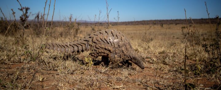
Pangolins look as though evolution got a little creative...covered in protective scales, equipped with long sticky tongues, and capable of curling into remarkably neat balls when threatened, these beautiful mammals seem to challenge many of the usual expectations for mammalian anatomy. Yet behind their distinctive appearance lies an evolutionary puzzle: where exactly do pangolins belong on the mammal family tree?
Pangolins and the Problem of Classification
Pangolins present a long-standing problem in classification. Due to their ant-eating diet, digging habits, and their uncanny appearance, they were historically grouped with animals such as anteaters and armadillos. Scientists now believe these similarities evolved independently as different species adapted over time to similar lifestyles, rather than them being related. In recent decades, molecular evidence has instead suggested that pangolins are the closest living relatives of the mammalian order Carnivora, the group that includes animals such as dogs and bears. Together, pangolins and carnivorans belong to a larger evolutionary Clade known as Ferae. The problem, however, is that there has been relatively little anatomical evidence to support this relationship.
To tackle this problem, an article published in the Zoological Journal of the Linnean Society addresses the lack of anatomical evidence linking pangolins with the Clade Ferae.
From Teeth to Skull: A Change in Focus
The study of evolutionary relationships between mammals, known as mammalian phylogenetics, is heavily reliant on teeth. Teeth often preserve well in the fossil record and contain features that help scientists identify relationships between species living millions of years apart. Unfortunately, pangolins never got the memo about how useful teeth are for classification. Being entirely toothless, researchers are left with far fewer of the usual clues to work with. As a result, scientists turned to a part of the body that is often overlooked in evolutionary studies: the skull.
Surprisingly, the network of blood vessels within the skull is not usually the first thing scientists look at when classifying mammals…
However, the veins that play a minor role in draining blood away from the brain, known as diploic veins, may be useful in unusual classification cases. As these veins run through the skull, they leave canals and grooves on the bone that can remain preserved in fossils. How convenient! Using this approach, researchers compared the diploic veins present in a wide range of placental species to investigate whether pangolins fit within a broader evolutionary pattern.
What Lies Beneath the Bone?
To investigate these hidden structures, researchers used micro-computed tomography (micro-CT) scanning alongside other imaging techniques to examine the internal anatomy of the skulls. The study analysed 92 specimens from 68 species, including species still living today and extinct species preserved in the fossil record, allowing a few long-deceased mammals to have their say in the discussion!
This is where some of the missing pieces of the puzzle begin to fit together. Pangolins possess an exceptionally dense network of diploic veins spread through the skull. Unlike the relatively modest diploic veins seen in most placental mammals, their diploic veins are far more developed and branch extensively through the cranial bones.
What makes this finding particularly interesting is not just the veins themselves but the role that they appear to play. In most mammals, a major vein, known as the superior sagittal sinus, is largely responsible for draining blood away from the brain after oxygen has been delivered. However, this study found that pangolins possess a reduced superior sagittal sinus, meaning that the enlarged diploic veins appear to take on a much greater role in this drainage system. This suggests that their unique venous network is not simply just a structural quirk, but part of a functional shift in how blood is transported away from the brain!
Extinct and Extant
It is important to note that this pattern was not limited to living pangolin species alone. Similar venous networks were identified in extinct pangolin species, suggesting that this feature has existed for a significant portion of pangolin history. The same general pattern was also observed in juvenile pangolin specimens, indicating that the pattern develops early in life and is unlikely to be an abnormality found only in adults.
The researchers also found that some carnivorans displayed partial similarities with pangolins in their cranial venous systems, although the networks were generally less extensive than those found in pangolins (Figure 1). Nevertheless, these similarities provide some of the long-sought anatomical evidence connecting pangolins with their closest living relatives, the carnivorans.
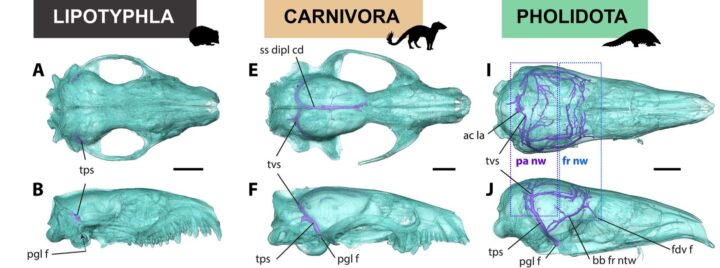
Figure 1: Comparison of cranial venous structures in the European hedgehog (A and B), carnivorans (E and F), and pangolin (I and J) skulls. Taken from Figure 1 of Billet et al., 2026.
Putting the Pieces Together
This study adds an intriguing piece of evidence to the pangolin puzzle. By examining diploic veins, the researchers introduced a form of evidence that has rarely been explored in mammalian phylogenetics.
Perhaps the most interesting takeaway is that sometimes the clues that we rely upon the most are not the ones that can tell us the full story. In the case of pangolins, focusing on teeth and more obvious skeletal features left major gaps in our understanding of their evolutionary relationships. Investigating the venous system within the skull gave us a few new pieces to the puzzle, offering a different perspective that may help to bridge the disconnect between anatomical and molecular evidence. Modern imaging techniques may also allow researchers to revisit overlooked anatomical structures.
For an animal that has spent decades confusing scientists, pangolins may finally be beginning to tell their own evolutionary story, one skull vein at a time.

Guest Blogger
Written by Shana Knight, a recent MSc graduate in Endangered Species Recovery and Conservation, with interests in applying quantitative ecological modelling and field-based research to biodiversity management, species recovery, climate change adaptation, and human–wildlife conflict mitigation. Edited by Georgia Cowie.
About the Journal
This blog was inspired by a paper published in our Zoological Journal, an international journal publishing high-quality papers covering systematic & evolutionary research from species both alive and extinct. Want to contribute to a blog? Contact the Journal Officer directly.